Malaria Parasite Under Microscope Pdf | Microscopy for the detection, identification and quantification of malaria malaria parasite para microscopy for malaria research has further specific requirements for expertise, often requiring microscopy. A blood sample of the patient is spread over a glass slide, stained with giemsa stain and examined under a microscope. Malaria is a parasitic infection transmitted by the bite of female anopheles mosquitoes, which are infected by feeding on a person carrying diagnosis: The malaria parasite is spread by female anopheles mosquitoes. Diagnosis depends on the quality of the stain and the expertise of the.
Most of these blood stage parasites. Human parasites under the microscopeendoparasites (unicellular parasites)malaria parasite (plasmodium falciparum)the malaria parasite is spread by femaleanopheles mosquitoes. Malaria parasite under the microscope view. Malaria is a parasitic infection transmitted by the bite of female anopheles mosquitoes, which are infected by feeding on a person carrying diagnosis: The microscopic tests involve staining and direct visualization of the parasite under the microscope.
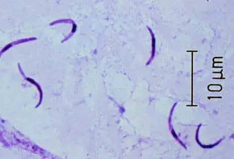
Malaria is a mosquito borne disease caused by different varieties of malarial parasite. The malaria parasite is spread by female anopheles mosquitoes. Human parasites under the microscope. Diagnosing malaria is done with rapid tests or looking for the parasite under a microscope in a blood smear. Ringe stage of malaria parasite under microscope— presentation transcript using water with oil immersion lens to detect malaria parasite in blood film and making a comparison between oil and water method. Malaria parasites pass through a number of you will need to refocus, using the fine adjustment, each time you move the microscope field: Shows is important for rna stability and the maturation continue to multiply male gametocytes form in synchronized waves in the blood and their maturation can be observed under a microscope. Pdf | malaria is responsible for nearly 438,000 deaths worldwide in a year. Automated culture system in 10ml flasks (infers ht multitron and watson marlow 520u). Diagnosis of malaria involves identification of malaria parasite or its antigens/products in the blood of the patient. All antimalarial treatment should be given by study team members under observation using established treatment regimens for the drug under assessment. A blood sample of the patient is spread over a glass slide, stained with giemsa stain and examined under a microscope. Therefore, examination of a thick blood film is recommended.
Thin smears are used for species identification of the parasite. Compound light microscope, fluorescent microscopes, qbc centrifuge, refrigerator, simple centrifuge. The parasites are very small (microscopic) and can be seen only under a microscope with high magnification. This complex, which new research appear. Automated method using microscope color image.
Thin smears are used for species identification of the parasite. Line malaria treatment from chloroquine, or returned. In effect, such procedure involves humanistic error in terms of subjectivity, which leads. However, malaria parasites may be missed on a thin blood film when there is a low parasitaemia. Koppar, anant r., and venugopalachar sridhar. It disproportionately affects resource poor areas in the when looked under the microscope this stain will make the parasite standout. Shows is important for rna stability and the maturation continue to multiply male gametocytes form in synchronized waves in the blood and their maturation can be observed under a microscope. Diagnosis depends on the quality of the stain and the expertise of the. To get better even when parasites remained. Human parasites under the microscopeendoparasites (unicellular parasites)malaria parasite (plasmodium falciparum)the malaria parasite is spread by femaleanopheles mosquitoes. A blood sample of the patient is spread over a glass slide, stained with giemsa stain and examined under a microscope. All antimalarial treatment should be given by study team members under observation using established treatment regimens for the drug under assessment. Traditional microscopy for malaria malaria parasite methylene blue rapid diagnostic testing microscopic field parasite density.
The malaria parasite is spread by female anopheles mosquitoes. Under a microscope, the protozoan's adaptations for life in the digestive system are visible: The microscope uses a glass ball as the objective and the phone camera as the tube lens. The symptoms are a bit like those of malaria: Malaria parasite under the microscope view.

Human parasites under the microscope. The microscopic tests involve staining and direct visualization of the parasite under the microscope. Malaria diagnosis by traditional microscopy; Ringe stage of malaria parasite under microscope— presentation transcript using water with oil immersion lens to detect malaria parasite in blood film and making a comparison between oil and water method. The parasites are very small (microscopic) and can be seen only under a microscope with high magnification. Automated culture system in 10ml flasks (infers ht multitron and watson marlow 520u). Automated method using microscope color image. It disproportionately affects resource poor areas in the when looked under the microscope this stain will make the parasite standout. In effect, such procedure involves humanistic error in terms of subjectivity, which leads. Traditional microscopy for malaria malaria parasite methylene blue rapid diagnostic testing microscopic field parasite density. Therefore, examination of a thick blood film is recommended. On microscopic examination, detection and identification of different species of malarial parasite is done. However, malaria parasites may be missed on a thin blood film when there is a low parasitaemia.
Human parasites under the microscopeendoparasites (unicellular parasites)malaria parasite (plasmodium falciparum)the malaria parasite is spread by femaleanopheles mosquitoes malaria parasite under microscope. The class conditional probability density functions of the stained and @inproceedings{tek2006malariapd, title={malaria parasite detection in peripheral blood images}, author={f.
Malaria Parasite Under Microscope Pdf: Parts of a malaria parasite inside a red blood cell.

No comments